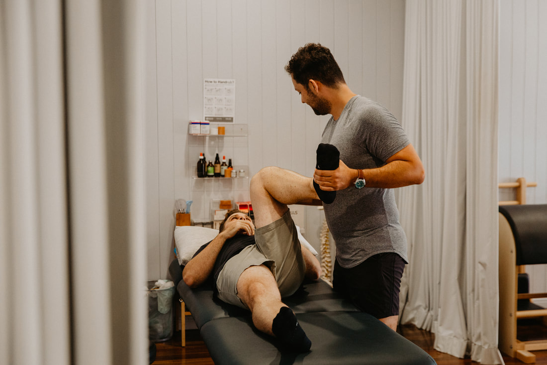
Ankle Osteoarthritis Physiotherapy Brisbane southside.
What is Ankle Osteoarthritis?
Ankle osteoarthritis, also known as degenerative arthritis of the ankle, is a joint condition characterised by the gradual breakdown of the cartilage that covers the ends of the bones in the ankle joint. This degeneration leads to pain, stiffness, and reduced joint mobility.
What causes Ankle Osteoarthritis?
Ankle osteoarthritis typically develops due to a combination of factors, including:
What are the symptoms of Ankle Osteoarthritis?
The most common symptoms of ankle osteoarthritis include:
How is Ankle Osteoarthritis diagnosed?
Ankle osteoarthritis is typically diagnosed through a combination of:
What is the treatment for Ankle Osteoarthritis?
The management of ankle osteoarthritis aims to alleviate pain, improve joint function, and enhance the patient's quality of life. Common treatment options include:
How is Ankle Osteoarthritis prevented?
While it may not be possible to prevent ankle osteoarthritis entirely, some preventive measures can help reduce the risk:
How can physiotherapy help with Ankle Osteoarthritis?
Physiotherapy can be a valuable part of the treatment plan for ankle osteoarthritis. Physiotherapists can help individuals with ankle osteoarthritis by addressing pain, improving joint function, and enhancing mobility. Here's how physiotherapy can help with ankle osteoarthritis:
If you or a loved one has questions about Ankle Osteoarthritis and how our physiotherapists might be able to help please call us on 07 3706 3407 or email [email protected]. We would love to work with you!
Ankle osteoarthritis, also known as degenerative arthritis of the ankle, is a joint condition characterised by the gradual breakdown of the cartilage that covers the ends of the bones in the ankle joint. This degeneration leads to pain, stiffness, and reduced joint mobility.
What causes Ankle Osteoarthritis?
Ankle osteoarthritis typically develops due to a combination of factors, including:
- Aging: The natural wear and tear on joints over time can lead to the development of osteoarthritis.
- Previous Injuries: A history of ankle injuries, such as sprains or fractures, can increase the risk of developing osteoarthritis in the ankle joint.
- Joint Overuse: Engaging in repetitive activities or putting excessive strain on the ankle joint can accelerate the breakdown of cartilage.
- Genetics: Some individuals may have a genetic predisposition to osteoarthritis.
What are the symptoms of Ankle Osteoarthritis?
The most common symptoms of ankle osteoarthritis include:
- Pain: Persistent pain in the ankle joint, especially during weight-bearing activities or movement.
- Stiffness: Reduced joint flexibility and difficulty moving the ankle.
- Swelling: Swelling and inflammation in the ankle joint.
- Decreased Range of Motion: Limited ability to move the ankle through its full range of motion.
- Tenderness: The ankle joint may be tender to the touch.
- Crepitus: A cracking or grating sensation may be felt or heard when moving the ankle.
How is Ankle Osteoarthritis diagnosed?
Ankle osteoarthritis is typically diagnosed through a combination of:
- Medical History: A healthcare provider will review your medical history, including any previous injuries or conditions that may contribute to ankle osteoarthritis.
- Physical Examination: A physical examination of the ankle joint will be conducted to assess the range of motion, tenderness, and stability.
- Imaging Studies: X-rays or other imaging tests, such as MRI or CT scans, may be ordered to visualise the extent of joint damage and the presence of bone spurs.
What is the treatment for Ankle Osteoarthritis?
The management of ankle osteoarthritis aims to alleviate pain, improve joint function, and enhance the patient's quality of life. Common treatment options include:
- Pain Management: Over-the-counter pain relievers or prescription medications may be recommended to manage pain and inflammation.
- Physical Therapy: A physical therapist can design an exercise program to strengthen the muscles around the ankle joint, improve joint stability, and enhance range of motion.
- Orthotic Devices: Custom-made or over-the-counter ankle braces, arch supports, or shoe inserts (orthotics) can provide added stability and reduce joint stress.
- Corticosteroid Injections: In some cases, injections of corticosteroids into the ankle joint can help reduce pain and inflammation.
- Lifestyle Modifications: Avoiding activities that exacerbate symptoms and maintaining a healthy weight can reduce the strain on the ankle joint.
- Assistive Devices: The use of canes or other assistive devices may be recommended to reduce the load on the affected ankle.
- Surgical Intervention: In severe cases of ankle osteoarthritis that do not respond to conservative treatments, surgical options such as ankle fusion or ankle replacement surgery may be considered.
How is Ankle Osteoarthritis prevented?
While it may not be possible to prevent ankle osteoarthritis entirely, some preventive measures can help reduce the risk:
- Avoid excessive strain on the ankle joint.
- Wear appropriate footwear with good support.
- Engage in regular exercise to maintain joint health and flexibility.
- Seek prompt treatment for ankle injuries to prevent long-term damage.
How can physiotherapy help with Ankle Osteoarthritis?
Physiotherapy can be a valuable part of the treatment plan for ankle osteoarthritis. Physiotherapists can help individuals with ankle osteoarthritis by addressing pain, improving joint function, and enhancing mobility. Here's how physiotherapy can help with ankle osteoarthritis:
- Assessment and Diagnosis: A physiotherapist will conduct a thorough assessment to evaluate the severity of your ankle osteoarthritis, including the extent of joint damage, pain levels, and functional limitations.
- Pain Management: Physiotherapists can employ various techniques to manage pain associated with ankle osteoarthritis, such as:
- Manual therapy: Hands-on techniques like joint mobilisation and soft tissue massage to reduce pain and improve joint mobility.
- Heat or cold therapy: Application of heat or cold packs to alleviate pain and inflammation.
- Ultrasound therapy or electrical stimulation: These modalities can help reduce pain and promote tissue healing.
- Range of Motion Exercises: Physiotherapists can design a customised exercise program to improve the range of motion in the ankle joint. Gentle, controlled exercises can help maintain or restore flexibility.
- Strengthening Exercises: Strengthening the muscles surrounding the ankle joint is essential for stability and support. Physiotherapists can provide exercises that target specific muscle groups to improve joint function.
- Balance and Proprioception Training: Ankle osteoarthritis can affect balance and proprioception (awareness of joint position). Physiotherapists can implement exercises and activities to improve balance and reduce the risk of falls.
- Gait Training: A physiotherapist can assess your walking pattern (gait) and provide guidance on modifications to reduce stress on the affected ankle.
- Orthotic Devices: Custom-made or over-the-counter orthotic insoles or braces may be recommended to provide added support, correct foot mechanics, and reduce joint strain.
- Footwear Recommendations: Physiotherapists can offer advice on appropriate footwear choices that provide stability and cushioning for the ankle joint.
- Assistive Devices: If necessary, physiotherapists can recommend the use of assistive devices, such as canes or crutches, to reduce pressure on the affected ankle during weight-bearing activities.
- Education: Physiotherapists provide education on proper joint protection techniques, lifestyle modifications, and exercises that can be continued at home to maintain joint health.
- Monitoring and Progress Evaluation: Throughout your physiotherapy treatment, your progress will be closely monitored, and adjustments to your exercise program or treatment plan will be made as needed to ensure you are on track to achieve your goals.
If you or a loved one has questions about Ankle Osteoarthritis and how our physiotherapists might be able to help please call us on 07 3706 3407 or email [email protected]. We would love to work with you!