Femoroacetabular Impingement Syndrome (FAI)
What is femoroacetabular impingement?
Femoroacetabular impingement syndrome (FAIS) is a common condition of the hip that can cause significant damage to the joint, leading to degeneration and osteoarthritis. FAIS constitutes an abnormal and dynamic contact between the femoral head-neck junction and the acetabular rim, resulting from altered bone morphology at one or both sites. Repetitive trauma at the site of impingement generates progressive damage to the acetabular labrum, chondrolabral junction, and articular cartilage.
FAI is most common in young to middle-aged active adults — particularly athletes in sports requiring repetitive hip flexion and rotation including football, hockey, ballet, cycling and martial arts — though it is not exclusively a sporting condition. It is one of the most frequently identified causes of hip and groin pain in active adults under 40.
Types of FAI
Three morphological types are recognised:
What are the symptoms?
Symptoms of FAI most often present as pain in the hip and groin region, typically when the hip is undergoing movement, as well as various degrees of restriction, noises such as clicking, locking, and stiffness. Pain is usually at the front of the hip and commonly provoked by hip flexion and internal rotation.
Deep groin pain — sometimes described as a "C-sign" where patients cup their hand around the lateral hip and anterior groin — with hip flexion and internal rotation is the characteristic clinical pattern. Prolonged sitting, getting in and out of a car, squatting and pivoting are common aggravating activities. Pain may also be felt in the anterior thigh, buttock or lateral hip.
How is it diagnosed?
The diagnosis is confirmed by the combination of clinical assessment — a detailed examination by a physiotherapist or orthopaedic specialist evaluating hip mobility, strength, and any movement that triggers pain — imaging including X-rays which can reveal abnormal bone shapes while MRI scans inspect damage to cartilage or labrum, and patient history understanding when the pain started, what aggravates it, and its nature.
The FADIR test — flexion, adduction and internal rotation — is the most sensitive clinical provocation test for FAI, reproducing the anterior hip pain by recreating the impingement contact. The FABER test assesses posterior hip and SIJ involvement. Hip strength testing — particularly hip abductor and external rotator strength — identifies the muscular deficits that contribute to symptoms and guide rehabilitation priorities.
Importantly, FAI morphology is present in a significant proportion of asymptomatic adults — particularly cam morphology in male athletes. The presence of bony changes on imaging must be interpreted alongside the clinical presentation to confirm FAI syndrome as the diagnosis.
Surgery versus conservative management
This is the most important question for most patients presenting with FAI, and the evidence has evolved significantly in recent years toward a more conservative-first approach.
Multiple randomised controlled trials and systematic reviews have now demonstrated that structured physiotherapy produces outcomes comparable to hip arthroscopy for FAI syndrome in many patients over twelve to twenty-four months. The FAIT trial — a randomised controlled trial comparing physiotherapy to hip arthroscopy — found no significant difference in outcomes at eight months, with both groups improving substantially from baseline. This does not mean surgery is never appropriate — it means a well-structured physiotherapy program should be the first approach for most patients with FAI syndrome, with surgery considered for those who fail conservative management or have large labral tears requiring repair.
How can physiotherapy help?
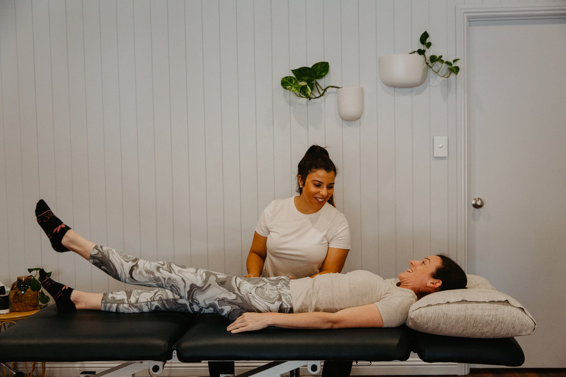
Physiotherapy for FAI involves exercise prescription developing a customised exercise program to strengthen hip muscles, improve joint stability, and address imbalances thereby reducing the impact of FAI. Range of motion improvement utilises targeted stretching and joint mobilisation to enhance hip joint flexibility and mitigate restrictions. Biomechanical correction involves a thorough assessment of movement patterns to identify and address abnormal mechanics contributing to FAI, helping to optimise hip function and minimise impingement.
The physiotherapy approach for FAI addresses both the structural impingement and the movement pattern contributors. Hip external rotator and abductor strengthening — the gluteus medius, gluteus minimus and deep external rotators — reduces the dynamic impingement that occurs when these muscles are weak and the femoral head migrates anteriorly during hip flexion. This is the most consistently impactful exercise intervention for FAI symptoms.
Lumbopelvic control — core stability, pelvic neutral positioning, and the movement coordination between the lumbar spine and hip — is equally important. Altered lumbopelvic mechanics place the hip in positions of greater impingement during functional activities, and correcting these patterns reduces the frequency and severity of impingement contact.
Activity modification during rehabilitation — identifying and temporarily avoiding the specific activities that repeatedly drive the hip into impingement — allows inflammation to settle while the strengthening program builds the capacity to tolerate loading again. This is not permanent avoidance but strategic load management during rehabilitation.
Real time ultrasound assists in retraining deep hip stabiliser activation. Clinical Pilates provides a controlled environment for progressive hip and lumbopelvic strengthening in positions that avoid the end-range impingement that provokes symptoms.
For patients who proceed to hip arthroscopy — including labral repair or debridement — structured post-surgical physiotherapy is central to achieving the best outcomes. See our hip pain page for the broader context of hip conditions.
Our physiotherapists Eliane Machado and Emma Cameron both have experience in hip conditions and FAI management and are members of the Australian Physiotherapy Association. Eliane's doctoral research in lower limb biomechanics is directly relevant to the movement analysis and hip mechanics assessment central to FAI rehabilitation.
To book or find out more, call us on 07 3706 3407 or book online below. We see patients from across Brisbane's southside including Tarragindi, Coorparoo, Holland Park, Greenslopes and Mt Gravatt.
Femoroacetabular impingement syndrome (FAIS) is a common condition of the hip that can cause significant damage to the joint, leading to degeneration and osteoarthritis. FAIS constitutes an abnormal and dynamic contact between the femoral head-neck junction and the acetabular rim, resulting from altered bone morphology at one or both sites. Repetitive trauma at the site of impingement generates progressive damage to the acetabular labrum, chondrolabral junction, and articular cartilage.
FAI is most common in young to middle-aged active adults — particularly athletes in sports requiring repetitive hip flexion and rotation including football, hockey, ballet, cycling and martial arts — though it is not exclusively a sporting condition. It is one of the most frequently identified causes of hip and groin pain in active adults under 40.
Types of FAI
Three morphological types are recognised:
- Cam impingement — where excess bone at the femoral head-neck junction creates a non-spherical femoral head. During hip flexion, this bony prominence contacts the acetabular rim and labrum, shearing the chondrolabral junction. Cam morphology is more common in males and in athletes who played high-load hip sports during adolescence — the bony prominence may develop as an adaptive response to loading during skeletal growth.
- Pincer impingement — where excess acetabular coverage (over-coverage of the femoral head by the acetabulum) causes the acetabular rim to impinge against the femoral head-neck junction during hip movement. Pincer morphology is more common in females.
- Combined impingement — where both cam and pincer morphology are present simultaneously, the most common presentation in clinical practice.
What are the symptoms?
Symptoms of FAI most often present as pain in the hip and groin region, typically when the hip is undergoing movement, as well as various degrees of restriction, noises such as clicking, locking, and stiffness. Pain is usually at the front of the hip and commonly provoked by hip flexion and internal rotation.
Deep groin pain — sometimes described as a "C-sign" where patients cup their hand around the lateral hip and anterior groin — with hip flexion and internal rotation is the characteristic clinical pattern. Prolonged sitting, getting in and out of a car, squatting and pivoting are common aggravating activities. Pain may also be felt in the anterior thigh, buttock or lateral hip.
How is it diagnosed?
The diagnosis is confirmed by the combination of clinical assessment — a detailed examination by a physiotherapist or orthopaedic specialist evaluating hip mobility, strength, and any movement that triggers pain — imaging including X-rays which can reveal abnormal bone shapes while MRI scans inspect damage to cartilage or labrum, and patient history understanding when the pain started, what aggravates it, and its nature.
The FADIR test — flexion, adduction and internal rotation — is the most sensitive clinical provocation test for FAI, reproducing the anterior hip pain by recreating the impingement contact. The FABER test assesses posterior hip and SIJ involvement. Hip strength testing — particularly hip abductor and external rotator strength — identifies the muscular deficits that contribute to symptoms and guide rehabilitation priorities.
Importantly, FAI morphology is present in a significant proportion of asymptomatic adults — particularly cam morphology in male athletes. The presence of bony changes on imaging must be interpreted alongside the clinical presentation to confirm FAI syndrome as the diagnosis.
Surgery versus conservative management
This is the most important question for most patients presenting with FAI, and the evidence has evolved significantly in recent years toward a more conservative-first approach.
Multiple randomised controlled trials and systematic reviews have now demonstrated that structured physiotherapy produces outcomes comparable to hip arthroscopy for FAI syndrome in many patients over twelve to twenty-four months. The FAIT trial — a randomised controlled trial comparing physiotherapy to hip arthroscopy — found no significant difference in outcomes at eight months, with both groups improving substantially from baseline. This does not mean surgery is never appropriate — it means a well-structured physiotherapy program should be the first approach for most patients with FAI syndrome, with surgery considered for those who fail conservative management or have large labral tears requiring repair.
How can physiotherapy help?
Physiotherapy for FAI involves exercise prescription developing a customised exercise program to strengthen hip muscles, improve joint stability, and address imbalances thereby reducing the impact of FAI. Range of motion improvement utilises targeted stretching and joint mobilisation to enhance hip joint flexibility and mitigate restrictions. Biomechanical correction involves a thorough assessment of movement patterns to identify and address abnormal mechanics contributing to FAI, helping to optimise hip function and minimise impingement.
The physiotherapy approach for FAI addresses both the structural impingement and the movement pattern contributors. Hip external rotator and abductor strengthening — the gluteus medius, gluteus minimus and deep external rotators — reduces the dynamic impingement that occurs when these muscles are weak and the femoral head migrates anteriorly during hip flexion. This is the most consistently impactful exercise intervention for FAI symptoms.
Lumbopelvic control — core stability, pelvic neutral positioning, and the movement coordination between the lumbar spine and hip — is equally important. Altered lumbopelvic mechanics place the hip in positions of greater impingement during functional activities, and correcting these patterns reduces the frequency and severity of impingement contact.
Activity modification during rehabilitation — identifying and temporarily avoiding the specific activities that repeatedly drive the hip into impingement — allows inflammation to settle while the strengthening program builds the capacity to tolerate loading again. This is not permanent avoidance but strategic load management during rehabilitation.
Real time ultrasound assists in retraining deep hip stabiliser activation. Clinical Pilates provides a controlled environment for progressive hip and lumbopelvic strengthening in positions that avoid the end-range impingement that provokes symptoms.
For patients who proceed to hip arthroscopy — including labral repair or debridement — structured post-surgical physiotherapy is central to achieving the best outcomes. See our hip pain page for the broader context of hip conditions.
Our physiotherapists Eliane Machado and Emma Cameron both have experience in hip conditions and FAI management and are members of the Australian Physiotherapy Association. Eliane's doctoral research in lower limb biomechanics is directly relevant to the movement analysis and hip mechanics assessment central to FAI rehabilitation.
To book or find out more, call us on 07 3706 3407 or book online below. We see patients from across Brisbane's southside including Tarragindi, Coorparoo, Holland Park, Greenslopes and Mt Gravatt.
Who to book in with:
Yulia Khasyanova
|
Emma Cameron
|
Eliane Machado
|