Spinal Stenosis Physiotherapy Brisbane southside.
What is Spinal Stenosis?
Spinal stenosis is a medical condition characterised by the narrowing of the spinal canal, which can put pressure on the spinal cord and the nerves that pass through it. This narrowing can occur in various regions of the spine and can lead to a range of symptoms.
What are the different types of Spinal Stenosis?
What causes Spinal Stenosis?
What are the symptoms of Spinal Stenosis?
How is a Spinal Stenosis diagnosed?
To diagnose spinal stenosis, a healthcare provider will typically perform a physical examination, review your medical history, and may order imaging tests such as X-rays, MRI (Magnetic Resonance Imaging), or CT (Computed Tomography) scans.
It's important to consult with a healthcare professional for a proper evaluation and treatment plan if you suspect you have spinal stenosis or are experiencing symptoms related to it. Treatment options should be discussed with your healthcare provider based on your specific condition and needs.
How can physiotherapy help with Spinal Stenosis?
Physiotherapy, also known as physical therapy, can be a valuable component of the treatment plan for individuals with spinal stenosis. While physiotherapy may not cure spinal stenosis, it can help manage symptoms, improve function, and enhance the overall quality of life for those affected. Here's how physiotherapy can be beneficial:
If you or a loved one has questions about Hip Stenosis and how our physiotherapists might be able to help please call us on 07 3706 3407 or email [email protected]. We would love to work with you!
Spinal stenosis is a medical condition characterised by the narrowing of the spinal canal, which can put pressure on the spinal cord and the nerves that pass through it. This narrowing can occur in various regions of the spine and can lead to a range of symptoms.
What are the different types of Spinal Stenosis?
- Lumbar Spinal Stenosis: This is the most common type and occurs in the lower back. It often affects older adults and can cause symptoms like lower back pain, leg pain, and numbness or weakness in the legs, particularly when walking or standing.
- Cervical Spinal Stenosis: This type occurs in the neck region of the spine and can lead to symptoms such as neck pain, shoulder pain, arm pain, and weakness or numbness in the arms and hands.
- Thoracic Spinal Stenosis: This is the least common type and affects the middle region of the spine. It can cause symptoms like chest pain, abdominal pain, and muscle weakness.
What causes Spinal Stenosis?
- Spinal stenosis is typically caused by age-related changes in the spine, such as the development of bone spurs, herniated discs, or thickened ligaments. These changes can narrow the spinal canal over time.
- In some cases, congenital factors or traumatic injuries can also contribute to spinal stenosis.
What are the symptoms of Spinal Stenosis?
- Symptoms of spinal stenosis can vary depending on the location and severity of the narrowing. Common symptoms include pain, numbness, tingling, and weakness in the affected area.
- Pain or discomfort often worsens with certain activities, such as walking or standing for extended periods, and may improve with rest or sitting.
How is a Spinal Stenosis diagnosed?
To diagnose spinal stenosis, a healthcare provider will typically perform a physical examination, review your medical history, and may order imaging tests such as X-rays, MRI (Magnetic Resonance Imaging), or CT (Computed Tomography) scans.
It's important to consult with a healthcare professional for a proper evaluation and treatment plan if you suspect you have spinal stenosis or are experiencing symptoms related to it. Treatment options should be discussed with your healthcare provider based on your specific condition and needs.
How can physiotherapy help with Spinal Stenosis?
Physiotherapy, also known as physical therapy, can be a valuable component of the treatment plan for individuals with spinal stenosis. While physiotherapy may not cure spinal stenosis, it can help manage symptoms, improve function, and enhance the overall quality of life for those affected. Here's how physiotherapy can be beneficial:
- Pain Management: Physiotherapists can use various techniques, such as manual therapy, modalities (e.g., heat or cold therapy), and electrotherapy, to help reduce pain associated with spinal stenosis. They may also teach you pain-relief strategies and exercises that you can do at home to alleviate discomfort.
- Posture and Body Mechanics: Physiotherapists can educate you on proper posture and body mechanics to reduce the strain on your spine. This includes teaching you how to sit, stand, lift, and perform daily activities with less stress on the affected area.
- Strengthening and Flexibility Exercises: Specific exercises prescribed by a physiotherapist can help strengthen the muscles that support the spine and improve flexibility. Strengthening the core muscles, in particular, can provide better spinal stability. Flexibility exercises can also help alleviate stiffness and increase the range of motion in the spine.
- Gait Training: If spinal stenosis affects your ability to walk or causes gait abnormalities, a physiotherapist can work with you on gait training exercises and techniques to improve your walking pattern.
- Balance and Coordination: Spinal stenosis can affect balance and coordination. Physiotherapy can include exercises to improve balance and reduce the risk of falls, especially for older individuals.
- Pain Relief Modalities: Physiotherapists may use modalities such as ultrasound, TENS (transcutaneous electrical nerve stimulation), or hot/cold therapy to help alleviate pain and reduce inflammation in the affected area.
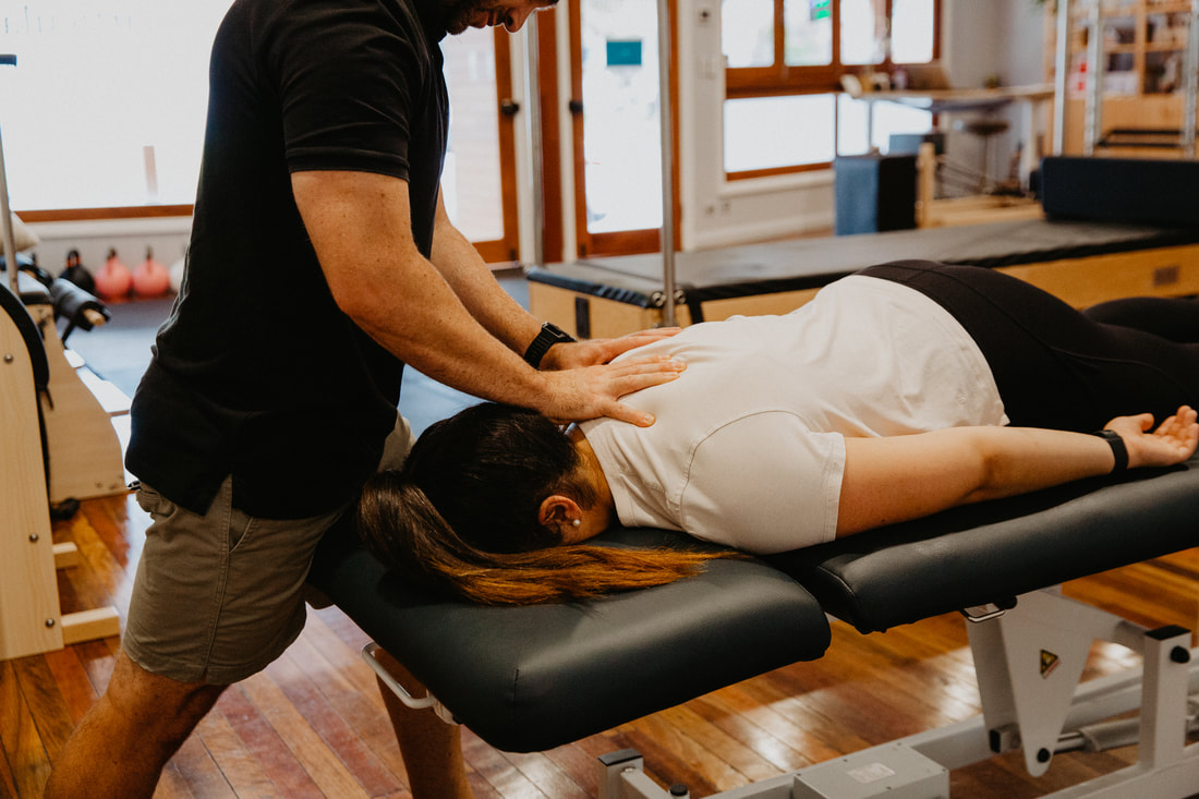
- Manual Therapy: Hands-on techniques like joint mobilisation or manipulation performed by a physiotherapist can help improve joint mobility and relieve pain.
- Education: Physiotherapists provide valuable education about the condition, including what activities to avoid and how to manage symptoms effectively. They can teach you ergonomic principles for your workplace or home environment.
- Assistive Devices: In some cases, a physiotherapist may recommend assistive devices like braces or orthotics to support the spine and improve mobility.
- Individualised Treatment Plans: One of the key benefits of physiotherapy is that treatment plans are tailored to each individual's specific needs and symptoms, taking into account the severity and location of spinal stenosis.
If you or a loved one has questions about Hip Stenosis and how our physiotherapists might be able to help please call us on 07 3706 3407 or email [email protected]. We would love to work with you!