Bursitis.
What is bursitis?
Bursitis is inflammation of a bursa — one of the small fluid-filled sacs distributed throughout the body at points of friction between bone, tendon, muscle and skin. Bursae act as cushions and lubricants, reducing friction at these high-movement interface points. When a bursa becomes irritated or inflamed, it fills with excess fluid, producing localised swelling, heat, pain and restricted movement at the affected joint.
Bursae exist at most major joints and several other anatomically vulnerable points — there are over 150 bursae in the body — but bursitis most commonly presents at the shoulder, hip, knee and elbow. The specific location of bursitis determines both its cause and its management, which is why site-specific assessment is important rather than treating all bursitis presentations identically.
What causes bursitis?
Bursitis develops through several distinct mechanisms. Overuse bursitis — the most common type — results from repetitive movements or sustained postures that create repetitive friction or compression over a bursa. Traumatic bursitis develops from a direct impact — a fall or blow directly over a superficial bursa. Inflammatory bursitis occurs secondary to systemic inflammatory conditions including rheumatoid arthritis, gout and psoriatic arthritis, where the inflammatory process directly involves the bursal tissue. Septic bursitis results from bacterial infection of the bursa — typically from a skin wound, puncture or haematogenous spread — and is a distinct condition requiring medical management with antibiotics.
Bursitis can occur around most joints in the body and causes considerable pain. If left untreated, prolonged symptoms can lead to tendon irritation causing broader damage. A physiotherapist can work with you to determine the best course of treatment, which often includes rest, cryotherapy, graded exposure to activity and correcting biomechanics.
Common sites of bursitis
Shoulder bursitis (subacromial bursitis) — inflammation of the subacromial bursa between the rotator cuff and the acromion — is the most common cause of shoulder pain in adults. It typically develops from repetitive overhead activity and is frequently associated with rotator cuff tendinopathy and shoulder impingement. See our shoulder bursitis page for the full clinical detail.
Hip bursitis (trochanteric bursitis) — inflammation of the bursa overlying the greater trochanter of the femur — produces characteristic lateral hip pain that is worse with lying on the affected side, stairs and prolonged walking. As discussed in detail on our hip bursitis page, isolated trochanteric bursitis is frequently associated with gluteal tendinopathy and greater trochanteric pain syndrome — distinguishing between these presentations guides the management approach.
Knee bursitis — several bursae around the knee can become inflamed. Prepatellar bursitis (over the kneecap) is common from repetitive kneeling and direct trauma. Infrapatellar bursitis (below the kneecap) is associated with patellar tendon overload. Pes anserine bursitis (medial knee) produces medial knee pain and is common in older adults with knee osteoarthritis. See our knee bursitis page for more detail.
Olecranon bursitis — inflammation of the bursa over the point of the elbow — produces a visible egg-shaped swelling at the posterior elbow. It is associated with repetitive leaning on the elbow (famously "student's elbow") and direct trauma. One third of olecranon bursitis cases are septic — requiring medical rather than physiotherapy management as the primary intervention.
Ischiogluteal bursitis — inflammation of the bursa over the ischial tuberosity — produces deep buttock pain that is exquisitely sensitive to prolonged sitting on hard surfaces. It is associated with cycling, rowing and sedentary occupations requiring prolonged hard-surface sitting.
Retrocalcaneal bursitis — between the Achilles tendon and the calcaneus — produces posterior heel pain and is frequently associated with Achilles tendinopathy, with which it can coexist.
What are the symptoms?
Bursitis presents as local swelling with possible heat and redness over the affected area and local stiffness. The swelling in superficial bursae — prepatellar, olecranon — is often clearly visible. Deeper bursae — subacromial, trochanteric — produce pain and restricted movement without visible swelling. Pain is typically well localised to the bursa site and is aggravated by direct pressure and by movements that compress or load the inflamed bursa.
How is it diagnosed?
Clinical assessment — including palpation of the bursa, assessment of the range of movement that reproduces symptoms, and specific provocative tests — is usually sufficient to diagnose bursitis. Ultrasound directly visualises bursal fluid and thickening and can guide aspiration or injection where indicated. MRI provides more comprehensive assessment of surrounding tendons and soft tissue, particularly where the diagnosis is uncertain or tendon pathology needs to be excluded.
A clinical point worth emphasising: bursitis does not cause sudden severe redness, warmth, fever or systemic illness — if these features are present, septic bursitis must be excluded urgently, as it requires antibiotic treatment and sometimes surgical drainage rather than physiotherapy.
How can physiotherapy help?
Physiotherapy is the primary treatment for non-septic bursitis across all common sites. The approach addresses both the acute bursal inflammation and the mechanical contributors — overuse patterns, muscle imbalances, biomechanical factors and training loads — that produced and perpetuate the condition.
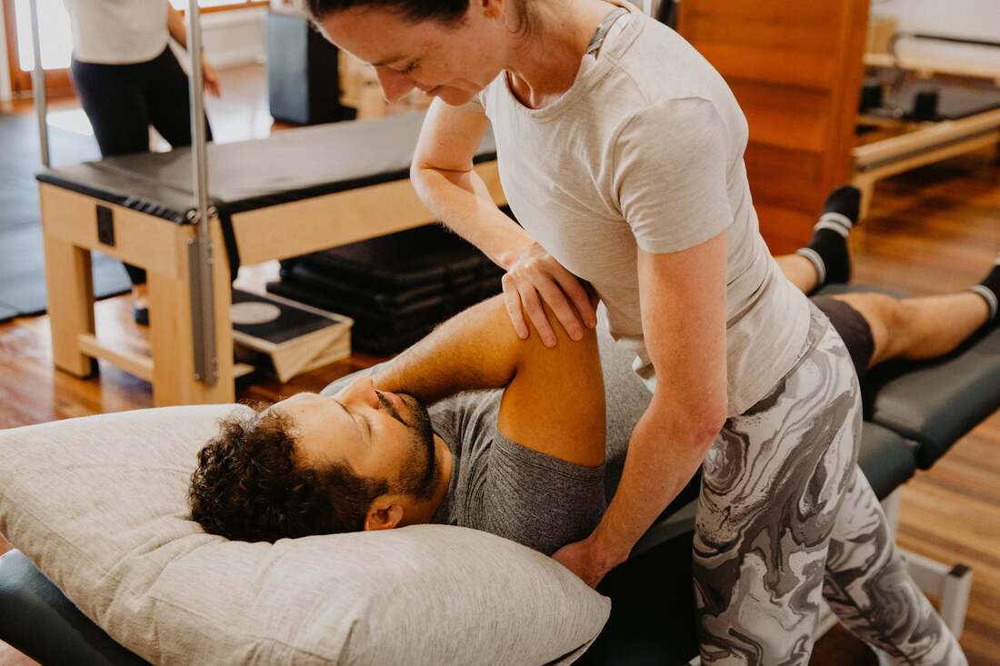
In the acute phase, activity modification to reduce the provocative loading, ice application and gentle movement within comfortable limits are the initial priorities. Manual therapy — soft tissue work and joint mobilisation of the relevant joint — reduces pain through neurophysiological mechanisms and addresses the secondary muscle guarding that develops around an inflamed bursa.
Progressive strengthening of the muscles surrounding the affected joint is the most important rehabilitation component — addressing the muscle imbalances and weakness that increase bursal loading during movement. Dry needling addresses myofascial trigger points in the muscles contributing to abnormal joint loading. Clinical Pilates provides a controlled environment for progressive strengthening. Real time ultrasound assists in retraining deep stabiliser activation.
Load management advice — workload modification, equipment adjustment, posture and technique correction — addresses the external contributors that will perpetuate bursitis if not modified alongside the clinical treatment. For workplace-related bursitis, WorkCover funded physiotherapy is available.
Our physiotherapists Bethany Kippen and Eliane Machado and Exercise Physiologist Ash O'Regan all have experience in bursitis management across all common sites and are members of the Australian Physiotherapy Association.
To book or find out more, call us on 07 3706 3407 or book online below. We see patients from across Brisbane's southside including Tarragindi, Coorparoo, Holland Park, Greenslopes and Mt Gravatt.
Bursitis is inflammation of a bursa — one of the small fluid-filled sacs distributed throughout the body at points of friction between bone, tendon, muscle and skin. Bursae act as cushions and lubricants, reducing friction at these high-movement interface points. When a bursa becomes irritated or inflamed, it fills with excess fluid, producing localised swelling, heat, pain and restricted movement at the affected joint.
Bursae exist at most major joints and several other anatomically vulnerable points — there are over 150 bursae in the body — but bursitis most commonly presents at the shoulder, hip, knee and elbow. The specific location of bursitis determines both its cause and its management, which is why site-specific assessment is important rather than treating all bursitis presentations identically.
What causes bursitis?
Bursitis develops through several distinct mechanisms. Overuse bursitis — the most common type — results from repetitive movements or sustained postures that create repetitive friction or compression over a bursa. Traumatic bursitis develops from a direct impact — a fall or blow directly over a superficial bursa. Inflammatory bursitis occurs secondary to systemic inflammatory conditions including rheumatoid arthritis, gout and psoriatic arthritis, where the inflammatory process directly involves the bursal tissue. Septic bursitis results from bacterial infection of the bursa — typically from a skin wound, puncture or haematogenous spread — and is a distinct condition requiring medical management with antibiotics.
Bursitis can occur around most joints in the body and causes considerable pain. If left untreated, prolonged symptoms can lead to tendon irritation causing broader damage. A physiotherapist can work with you to determine the best course of treatment, which often includes rest, cryotherapy, graded exposure to activity and correcting biomechanics.
Common sites of bursitis
Shoulder bursitis (subacromial bursitis) — inflammation of the subacromial bursa between the rotator cuff and the acromion — is the most common cause of shoulder pain in adults. It typically develops from repetitive overhead activity and is frequently associated with rotator cuff tendinopathy and shoulder impingement. See our shoulder bursitis page for the full clinical detail.
Hip bursitis (trochanteric bursitis) — inflammation of the bursa overlying the greater trochanter of the femur — produces characteristic lateral hip pain that is worse with lying on the affected side, stairs and prolonged walking. As discussed in detail on our hip bursitis page, isolated trochanteric bursitis is frequently associated with gluteal tendinopathy and greater trochanteric pain syndrome — distinguishing between these presentations guides the management approach.
Knee bursitis — several bursae around the knee can become inflamed. Prepatellar bursitis (over the kneecap) is common from repetitive kneeling and direct trauma. Infrapatellar bursitis (below the kneecap) is associated with patellar tendon overload. Pes anserine bursitis (medial knee) produces medial knee pain and is common in older adults with knee osteoarthritis. See our knee bursitis page for more detail.
Olecranon bursitis — inflammation of the bursa over the point of the elbow — produces a visible egg-shaped swelling at the posterior elbow. It is associated with repetitive leaning on the elbow (famously "student's elbow") and direct trauma. One third of olecranon bursitis cases are septic — requiring medical rather than physiotherapy management as the primary intervention.
Ischiogluteal bursitis — inflammation of the bursa over the ischial tuberosity — produces deep buttock pain that is exquisitely sensitive to prolonged sitting on hard surfaces. It is associated with cycling, rowing and sedentary occupations requiring prolonged hard-surface sitting.
Retrocalcaneal bursitis — between the Achilles tendon and the calcaneus — produces posterior heel pain and is frequently associated with Achilles tendinopathy, with which it can coexist.
What are the symptoms?
Bursitis presents as local swelling with possible heat and redness over the affected area and local stiffness. The swelling in superficial bursae — prepatellar, olecranon — is often clearly visible. Deeper bursae — subacromial, trochanteric — produce pain and restricted movement without visible swelling. Pain is typically well localised to the bursa site and is aggravated by direct pressure and by movements that compress or load the inflamed bursa.
How is it diagnosed?
Clinical assessment — including palpation of the bursa, assessment of the range of movement that reproduces symptoms, and specific provocative tests — is usually sufficient to diagnose bursitis. Ultrasound directly visualises bursal fluid and thickening and can guide aspiration or injection where indicated. MRI provides more comprehensive assessment of surrounding tendons and soft tissue, particularly where the diagnosis is uncertain or tendon pathology needs to be excluded.
A clinical point worth emphasising: bursitis does not cause sudden severe redness, warmth, fever or systemic illness — if these features are present, septic bursitis must be excluded urgently, as it requires antibiotic treatment and sometimes surgical drainage rather than physiotherapy.
How can physiotherapy help?
Physiotherapy is the primary treatment for non-septic bursitis across all common sites. The approach addresses both the acute bursal inflammation and the mechanical contributors — overuse patterns, muscle imbalances, biomechanical factors and training loads — that produced and perpetuate the condition.
In the acute phase, activity modification to reduce the provocative loading, ice application and gentle movement within comfortable limits are the initial priorities. Manual therapy — soft tissue work and joint mobilisation of the relevant joint — reduces pain through neurophysiological mechanisms and addresses the secondary muscle guarding that develops around an inflamed bursa.
Progressive strengthening of the muscles surrounding the affected joint is the most important rehabilitation component — addressing the muscle imbalances and weakness that increase bursal loading during movement. Dry needling addresses myofascial trigger points in the muscles contributing to abnormal joint loading. Clinical Pilates provides a controlled environment for progressive strengthening. Real time ultrasound assists in retraining deep stabiliser activation.
Load management advice — workload modification, equipment adjustment, posture and technique correction — addresses the external contributors that will perpetuate bursitis if not modified alongside the clinical treatment. For workplace-related bursitis, WorkCover funded physiotherapy is available.
Our physiotherapists Bethany Kippen and Eliane Machado and Exercise Physiologist Ash O'Regan all have experience in bursitis management across all common sites and are members of the Australian Physiotherapy Association.
To book or find out more, call us on 07 3706 3407 or book online below. We see patients from across Brisbane's southside including Tarragindi, Coorparoo, Holland Park, Greenslopes and Mt Gravatt.
Who to book in with:
Ash O'Regan
|
Emma Cameron
|
Bethany Kippen
|