Glenohumeral Joint Instability
What is glenohumeral joint instability?
The glenohumeral joint — the ball and socket joint between the humeral head and the glenoid of the scapula — is the most mobile joint in the human body. This extraordinary range of motion comes at a cost: the shoulder is inherently dependent on its surrounding soft tissue structures for stability in a way that more constrained joints like the hip are not. The labrum (a fibrocartilaginous rim that deepens the glenoid socket), the glenohumeral ligaments, the joint capsule and the rotator cuff muscles all work in concert to keep the humeral head centred in the glenoid during movement.
Glenohumeral instability occurs when this stabilising system is insufficient to maintain normal joint mechanics, producing a spectrum of problems from subtle excessive translation of the humeral head to frank dislocation where the ball leaves the socket entirely.
Types of glenohumeral instability
Understanding the type of instability is critical to selecting the right management approach — the treatment for traumatic anterior instability following a dislocation is quite different to the management of atraumatic multidirectional instability in a hypermobile gymnast.
Traumatic instability results from a specific injury — typically a shoulder dislocation or significant subluxation — that damages the static stabilisers. Anterior traumatic instability is the most common, usually resulting from a forced abduction and external rotation mechanism (falling on an outstretched arm or a tackle in contact sport). The characteristic structural injury is a Bankart lesion — detachment of the labrum from the anterior glenoid — sometimes associated with a Hill-Sachs lesion (a compression fracture of the posterior humeral head from impacting the glenoid rim during dislocation). The risk of recurrence after a first-time traumatic dislocation is significantly influenced by age — young active patients under 25 have recurrence rates approaching 90% without surgical intervention.
Atraumatic instability develops without a significant injury, from a combination of capsular laxity, muscle imbalance and repetitive microtrauma. It is more common in overhead athletes (swimmers, throwers, volleyball players) and in people with generalised joint hypermobility or Ehlers-Danlos syndrome. It typically presents as a vague sense of shoulder instability, apprehension with certain movements, and pain rather than frank dislocation.
Multidirectional instability (MDI) involves excessive laxity in multiple directions — anterior, posterior and inferior — and is almost exclusively atraumatic in origin. It is particularly associated with hypermobility conditions and overhead sport, and responds well to a structured rehabilitation program in most cases.
Posterior instability, while less common than anterior instability, is underdiagnosed and often missed. It is associated with contact sport (particularly rugby), weightlifting, and repetitive posterior loading. The apprehension and relocation tests used for anterior instability are not useful for posterior instability, and awareness of the different examination findings is important.
What are the symptoms?
Symptoms range from frank dislocations — where the shoulder visibly deforms and requires manual reduction — to subtle feelings of the shoulder slipping, clicking or feeling unreliable with certain movements. Apprehension — a sense of anxiety or muscular guarding when the shoulder is moved into positions that risk instability — is a characteristic and diagnostically important symptom. Pain, weakness, and avoidance of overhead or rotational activities are common functional consequences.
In hypermobility-related instability, patients often describe the shoulder as feeling "loose" or "not quite right" without clear episodes of dislocation, and may have developed habitual muscle patterns to guard against instability that produce secondary pain and dysfunction of their own.
How is it diagnosed?
Clinical assessment involves specific instability tests — the apprehension, relocation and surprise tests for anterior instability, load and shift testing, and the sulcus sign for inferior laxity — interpreted in the context of the history and functional presentation. Examination of both shoulders for comparison and assessment of generalised hypermobility is important.
MRI arthrogram — where contrast is injected into the shoulder joint before MRI — is the most sensitive imaging for labral pathology and capsular injuries. Standard MRI can miss subtle labral lesions. CT scan with 3D reconstruction is used for surgical planning in cases with significant bony defects.
How can physiotherapy help?
For atraumatic and multidirectional instability, physiotherapy is the primary and most effective treatment. The evidence for rotator cuff and periscapular strengthening programs in this population is strong, and the majority of patients achieve good to excellent outcomes without surgery.
The rehabilitation focus is on building the dynamic stabilisers of the shoulder — particularly the rotator cuff's role in compressing and centring the humeral head in the glenoid during movement. The subscapularis, infraspinatus and teres minor, along with the lower trapezius and serratus anterior for scapular control, are the primary targets. Proprioception and neuromuscular control training — teaching the shoulder's reflex stabilisation system to respond rapidly to perturbations — is equally important and is often neglected in generic shoulder programs.
For traumatic anterior instability following a first dislocation, the management decision depends significantly on age, activity level, and the structural findings on imaging. Young active patients have a high recurrence risk and many will ultimately require surgical stabilisation — but physiotherapy in the acute phase manages pain, restores range of motion, and builds rotator cuff function that improves outcomes whether the eventual pathway is surgical or conservative.
For post-surgical instability repair — including Bankart repair and Latarjet procedure — physiotherapy follows a structured protocol respecting the specific precautions of the surgical technique, progressing from early motion through to full strength and sport-specific rehabilitation.
For patients with hypermobility or EDS, the approach is more conservative in loading parameters and more comprehensive in scope — addressing the whole upper limb kinetic chain, including cervical and thoracic spine control, rather than the shoulder in isolation.
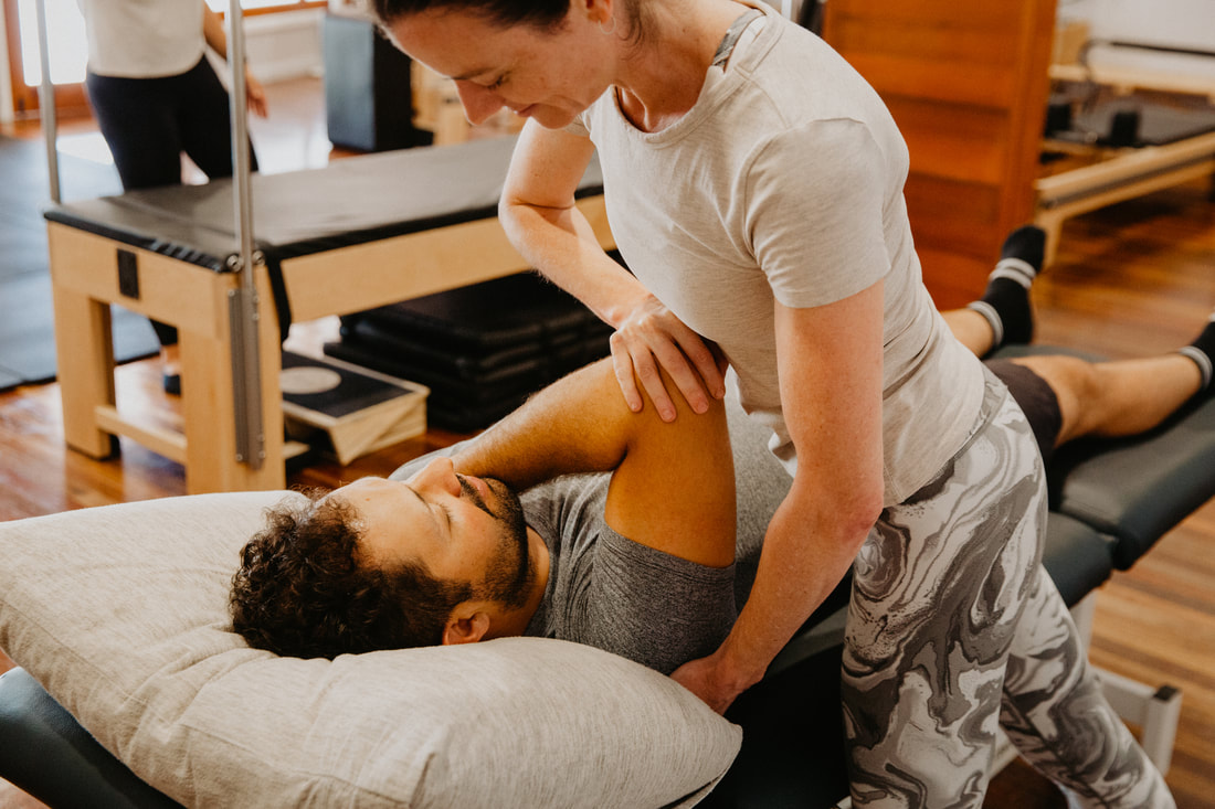
Clinical Pilates is an excellent tool for shoulder instability rehabilitation — the controlled environment allows progressive rotator cuff and scapular loading through ranges that can be precisely managed, building strength and neuromuscular control without exposing the shoulder to destabilising positions. Real time ultrasound assists in retraining deep rotator cuff activation where pain and inhibition are affecting muscle recruitment.
Our physiotherapists Bethany Kippen, Yulia Khasyanova and Emma Cameron all have experience in shoulder instability management and are members of the Australian Physiotherapy Association. Yulia's specialist background in hypermobility and connective tissue disorders is particularly relevant for patients with hypermobility-related shoulder instability.
To book or find out more, call us on 07 3706 3407 or book online below. We see patients from across Brisbane's southside including Tarragindi, Coorparoo, Holland Park, Greenslopes and Mt Gravatt.
The glenohumeral joint — the ball and socket joint between the humeral head and the glenoid of the scapula — is the most mobile joint in the human body. This extraordinary range of motion comes at a cost: the shoulder is inherently dependent on its surrounding soft tissue structures for stability in a way that more constrained joints like the hip are not. The labrum (a fibrocartilaginous rim that deepens the glenoid socket), the glenohumeral ligaments, the joint capsule and the rotator cuff muscles all work in concert to keep the humeral head centred in the glenoid during movement.
Glenohumeral instability occurs when this stabilising system is insufficient to maintain normal joint mechanics, producing a spectrum of problems from subtle excessive translation of the humeral head to frank dislocation where the ball leaves the socket entirely.
Types of glenohumeral instability
Understanding the type of instability is critical to selecting the right management approach — the treatment for traumatic anterior instability following a dislocation is quite different to the management of atraumatic multidirectional instability in a hypermobile gymnast.
Traumatic instability results from a specific injury — typically a shoulder dislocation or significant subluxation — that damages the static stabilisers. Anterior traumatic instability is the most common, usually resulting from a forced abduction and external rotation mechanism (falling on an outstretched arm or a tackle in contact sport). The characteristic structural injury is a Bankart lesion — detachment of the labrum from the anterior glenoid — sometimes associated with a Hill-Sachs lesion (a compression fracture of the posterior humeral head from impacting the glenoid rim during dislocation). The risk of recurrence after a first-time traumatic dislocation is significantly influenced by age — young active patients under 25 have recurrence rates approaching 90% without surgical intervention.
Atraumatic instability develops without a significant injury, from a combination of capsular laxity, muscle imbalance and repetitive microtrauma. It is more common in overhead athletes (swimmers, throwers, volleyball players) and in people with generalised joint hypermobility or Ehlers-Danlos syndrome. It typically presents as a vague sense of shoulder instability, apprehension with certain movements, and pain rather than frank dislocation.
Multidirectional instability (MDI) involves excessive laxity in multiple directions — anterior, posterior and inferior — and is almost exclusively atraumatic in origin. It is particularly associated with hypermobility conditions and overhead sport, and responds well to a structured rehabilitation program in most cases.
Posterior instability, while less common than anterior instability, is underdiagnosed and often missed. It is associated with contact sport (particularly rugby), weightlifting, and repetitive posterior loading. The apprehension and relocation tests used for anterior instability are not useful for posterior instability, and awareness of the different examination findings is important.
What are the symptoms?
Symptoms range from frank dislocations — where the shoulder visibly deforms and requires manual reduction — to subtle feelings of the shoulder slipping, clicking or feeling unreliable with certain movements. Apprehension — a sense of anxiety or muscular guarding when the shoulder is moved into positions that risk instability — is a characteristic and diagnostically important symptom. Pain, weakness, and avoidance of overhead or rotational activities are common functional consequences.
In hypermobility-related instability, patients often describe the shoulder as feeling "loose" or "not quite right" without clear episodes of dislocation, and may have developed habitual muscle patterns to guard against instability that produce secondary pain and dysfunction of their own.
How is it diagnosed?
Clinical assessment involves specific instability tests — the apprehension, relocation and surprise tests for anterior instability, load and shift testing, and the sulcus sign for inferior laxity — interpreted in the context of the history and functional presentation. Examination of both shoulders for comparison and assessment of generalised hypermobility is important.
MRI arthrogram — where contrast is injected into the shoulder joint before MRI — is the most sensitive imaging for labral pathology and capsular injuries. Standard MRI can miss subtle labral lesions. CT scan with 3D reconstruction is used for surgical planning in cases with significant bony defects.
How can physiotherapy help?
For atraumatic and multidirectional instability, physiotherapy is the primary and most effective treatment. The evidence for rotator cuff and periscapular strengthening programs in this population is strong, and the majority of patients achieve good to excellent outcomes without surgery.
The rehabilitation focus is on building the dynamic stabilisers of the shoulder — particularly the rotator cuff's role in compressing and centring the humeral head in the glenoid during movement. The subscapularis, infraspinatus and teres minor, along with the lower trapezius and serratus anterior for scapular control, are the primary targets. Proprioception and neuromuscular control training — teaching the shoulder's reflex stabilisation system to respond rapidly to perturbations — is equally important and is often neglected in generic shoulder programs.
For traumatic anterior instability following a first dislocation, the management decision depends significantly on age, activity level, and the structural findings on imaging. Young active patients have a high recurrence risk and many will ultimately require surgical stabilisation — but physiotherapy in the acute phase manages pain, restores range of motion, and builds rotator cuff function that improves outcomes whether the eventual pathway is surgical or conservative.
For post-surgical instability repair — including Bankart repair and Latarjet procedure — physiotherapy follows a structured protocol respecting the specific precautions of the surgical technique, progressing from early motion through to full strength and sport-specific rehabilitation.
For patients with hypermobility or EDS, the approach is more conservative in loading parameters and more comprehensive in scope — addressing the whole upper limb kinetic chain, including cervical and thoracic spine control, rather than the shoulder in isolation.
Clinical Pilates is an excellent tool for shoulder instability rehabilitation — the controlled environment allows progressive rotator cuff and scapular loading through ranges that can be precisely managed, building strength and neuromuscular control without exposing the shoulder to destabilising positions. Real time ultrasound assists in retraining deep rotator cuff activation where pain and inhibition are affecting muscle recruitment.
Our physiotherapists Bethany Kippen, Yulia Khasyanova and Emma Cameron all have experience in shoulder instability management and are members of the Australian Physiotherapy Association. Yulia's specialist background in hypermobility and connective tissue disorders is particularly relevant for patients with hypermobility-related shoulder instability.
To book or find out more, call us on 07 3706 3407 or book online below. We see patients from across Brisbane's southside including Tarragindi, Coorparoo, Holland Park, Greenslopes and Mt Gravatt.
Who to book in with:
Yulia Khasyanova
|
Bethany Kippen
|
Emma Cameron
|