Lateral Collateral Ligament (LCL) Injuries physiotherapy Brisbane southside.
What is a Lateral Collateral Ligament Injury?
The lateral collateral ligament (LCL) is one of the four major ligaments that stabilise the knee joint. It runs along the outside of the knee and helps to prevent excessive side-to-side movement. LCL injuries can occur when there is a force applied to the inside of the knee, pushing it outward, or when the knee is hyperextended. Common causes include sports-related injuries (e.g., football, soccer, skiing) and motor vehicle accidents. Hyperextension of the knee can also lead to LCL injuries.
What are the symptoms of a Lateral Collateral Ligament Injury?
The symptoms of an LCL injury may include pain along the outside of the knee, swelling, instability or a feeling of the knee giving way, and difficulty bending or straightening the knee. In more severe cases, there may be bruising or a noticeable deformity.
What are the different grades of a Lateral Collateral Ligament Injury?
LCL injuries are often graded based on their severity:
How is a Lateral Collateral Ligament Injury diagnosed?
A healthcare provider will typically perform a physical examination, assess the range of motion, and may order imaging tests such as X-rays or MRI scans to determine the extent of the injury.
What is the recovery time for a Lateral Collateral Ligament Injury?
Recovery time varies depending on the severity of the injury and the chosen treatment method. Grade 1 injuries may heal in a few weeks, while Grade 3 injuries may require several months of rehabilitation.
How can physiotherapy help with a Lateral Collateral Ligament Injury?
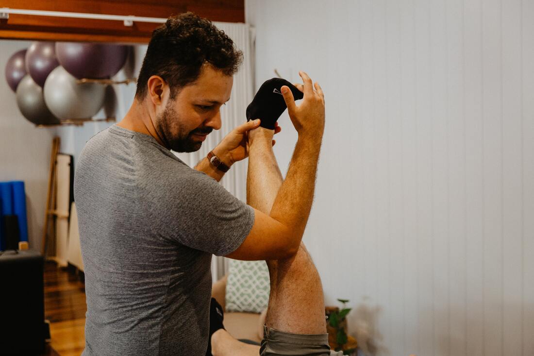
Physiotherapy plays a crucial role in the management and rehabilitation of lateral collateral ligament (LCL) injuries. A physiotherapist can design a customised treatment plan to help patients recover from LCL injuries, whether they are mild (Grade 1) or more severe (Grade 2 or 3). Here's how physiotherapy can be beneficial:
If you or a loved one has questions about a Lateral Collateral Ligament Injury and how our physiotherapists might be able to help please call us on 07 3706 3407 or email [email protected]. We would love to work with you!
The lateral collateral ligament (LCL) is one of the four major ligaments that stabilise the knee joint. It runs along the outside of the knee and helps to prevent excessive side-to-side movement. LCL injuries can occur when there is a force applied to the inside of the knee, pushing it outward, or when the knee is hyperextended. Common causes include sports-related injuries (e.g., football, soccer, skiing) and motor vehicle accidents. Hyperextension of the knee can also lead to LCL injuries.
What are the symptoms of a Lateral Collateral Ligament Injury?
The symptoms of an LCL injury may include pain along the outside of the knee, swelling, instability or a feeling of the knee giving way, and difficulty bending or straightening the knee. In more severe cases, there may be bruising or a noticeable deformity.
What are the different grades of a Lateral Collateral Ligament Injury?
LCL injuries are often graded based on their severity:
- Grade 1: Mild stretching or microscopic tears in the ligament.
- Grade 2: Partial tearing of the ligament.
- Grade 3: Complete tear of the ligament.
How is a Lateral Collateral Ligament Injury diagnosed?
A healthcare provider will typically perform a physical examination, assess the range of motion, and may order imaging tests such as X-rays or MRI scans to determine the extent of the injury.
What is the recovery time for a Lateral Collateral Ligament Injury?
Recovery time varies depending on the severity of the injury and the chosen treatment method. Grade 1 injuries may heal in a few weeks, while Grade 3 injuries may require several months of rehabilitation.
How can physiotherapy help with a Lateral Collateral Ligament Injury?
Physiotherapy plays a crucial role in the management and rehabilitation of lateral collateral ligament (LCL) injuries. A physiotherapist can design a customised treatment plan to help patients recover from LCL injuries, whether they are mild (Grade 1) or more severe (Grade 2 or 3). Here's how physiotherapy can be beneficial:
- Pain Management: Physiotherapists can use various techniques, such as manual therapy, modalities like ice or heat, and electrical stimulation, to help manage pain and reduce inflammation in the early stages of recovery.
- Reducing Swelling: Swelling is a common symptom of LCL injuries. Physiotherapists can employ techniques like ice and compression to reduce swelling, which can, in turn, alleviate pain and improve the healing process.
- Restoring Range of Motion: After an LCL injury, the knee may become stiff and lose its normal range of motion. Physiotherapy includes exercises and stretches to gradually restore the knee's flexibility and mobility.
- Strengthening Muscles: Weakness in the muscles around the knee can contribute to instability. Physiotherapists will design a strengthening program tailored to the individual's needs, focusing on the quadriceps, hamstrings, calf muscles, and hip muscles. Strengthening these muscles can provide better support to the knee joint.
- Improving Proprioception and Balance: LCL injuries can affect proprioception (awareness of the joint's position in space) and balance. Physiotherapy can include exercises to enhance proprioception and improve balance, reducing the risk of future injuries.
- Functional Rehabilitation: As the knee heals, physiotherapists introduce functional exercises and activities that mimic real-life movements. This helps patients regain confidence and prepare for a return to sports or daily activities.
- Bracing and Taping: In some cases, physiotherapists may use knee braces or taping techniques to provide additional support and stability to the injured knee during the rehabilitation process.
- Patient Education: Physiotherapists educate patients about their condition, the importance of adherence to the rehabilitation program, and strategies to prevent re-injury. They can also provide advice on proper biomechanics and techniques for activities that may have contributed to the injury.
- Progress Monitoring: Throughout the rehabilitation process, physiotherapists continuously assess progress and make necessary adjustments to the treatment plan. They may modify exercises and activities as the patient's condition improves.
- Return to Activity: The ultimate goal of physiotherapy is to safely return the patient to their desired level of activity, whether it's recreational sports, work, or daily life. Physiotherapists work with patients to ensure they have regained the strength, stability, and confidence needed for a safe return to their activities.
If you or a loved one has questions about a Lateral Collateral Ligament Injury and how our physiotherapists might be able to help please call us on 07 3706 3407 or email [email protected]. We would love to work with you!