Shoulder Bursitis.
What is shoulder bursitis?
Shoulder bursitis is inflammation of one of the bursae around the shoulder joint — most commonly the subacromial bursa, which sits between the rotator cuff tendons and the undersurface of the acromion (the bony roof of the shoulder). When this bursa becomes irritated or inflamed it fills with excess fluid, producing pain, swelling and restricted shoulder movement that is typically worst with overhead activity, reaching and lying on the affected side.
Shoulder bursitis is one of the most common causes of shoulder pain in adults — the subacromial space is a narrow anatomical corridor and any factor that reduces its dimensions or increases the load on the structures within it can produce bursal irritation. It is closely related to shoulder impingement syndrome and rotator cuff tendinopathy — in practice these conditions frequently coexist, and differentiating between them requires a careful clinical assessment rather than assuming the imaging diagnosis tells the whole story.
The subacromial space — why it matters
The subacromial space is the gap between the humeral head below and the acromion and coracoacromial ligament above, through which the supraspinatus tendon and subacromial bursa must pass during shoulder elevation. In a healthy shoulder, the rotator cuff muscles — particularly the supraspinatus — depress the humeral head during elevation, maintaining adequate subacromial space. When rotator cuff function is compromised from weakness, fatigue or inhibition, the humeral head rides higher in the socket, narrowing the subacromial space and increasing mechanical contact on the bursa and tendon.
This dynamic narrowing is the central mechanism of most subacromial bursitis presentations — it is a functional problem driven by rotator cuff and scapular muscle imbalance, not simply an anatomical problem with the shape of the acromion. This understanding is clinically important because it means physiotherapy addressing rotator cuff function and scapular control directly addresses the mechanism driving the bursitis — not just managing the symptoms.
What causes shoulder bursitis?
Several factors contribute to subacromial bursitis. Rotator cuff weakness or dysfunction — from overuse, injury or disuse — reduces the dynamic depression of the humeral head during shoulder movement. Poor scapular control — weak lower trapezius and serratus anterior — produces inadequate acromial elevation during shoulder elevation, reducing the subacromial space. Repetitive overhead activity in occupational or sporting contexts — painting, swimming, throwing, racquet sports — creates sustained mechanical loading on the bursa.
Postural factors — forward head posture, thoracic kyphosis, rounded shoulders — alter shoulder mechanics and reduce subacromial space.
Inflammatory conditions including rheumatoid arthritis and crystal arthropathy directly cause bursal inflammation. Acromioclavicular joint osteoarthritis produces inferior osteophytes that mechanically impinge on the subacromial contents. Direct trauma — a fall onto the outstretched hand or onto the point of the shoulder — can cause acute bursal haemorrhage and inflammation.
What are the symptoms?
Pain that worsens with overhead movements or reaching is the hallmark. A painful arc — where shoulder elevation is comfortable from 0 to 60 degrees, painful from 60 to 120 degrees, and comfortable again above 120 degrees — is the classic clinical finding of subacromial impingement and bursitis. Pain is typically located in the lateral deltoid region and anterior shoulder, and is often worse at night, particularly when lying on the affected side.
Stiffness and weakness with sustained overhead or reaching activities, and pain with reaching across the body — the impingement test — are consistent features. Referred pain into the lateral arm is common from both the inflamed bursa and the associated rotator cuff tendinopathy.
How is it diagnosed?
Diagnosis of shoulder bursitis typically involves a physical examination by a healthcare provider, along with a review of your medical history. Imaging tests like X-rays or ultrasound may be used to rule out other shoulder conditions.
Clinical assessment includes the Neer and Hawkins-Kennedy impingement tests, rotator cuff strength testing, scapular assessment, and the painful arc assessment. Ultrasound directly visualises bursal fluid and thickening, identifies rotator cuff tears, and can guide diagnostic or therapeutic corticosteroid injection. MRI provides the most comprehensive assessment of the rotator cuff, bursa and surrounding structures.
A diagnostic injection of local anaesthetic into the subacromial space — producing temporary relief of symptoms — confirms the subacromial origin of pain and assists in differentiating bursitis from glenohumeral joint or acromioclavicular joint sources.
How can physiotherapy help?
Physiotherapy is the primary treatment for subacromial bursitis and addresses both the acute inflammation and the underlying biomechanical contributors that produced and perpetuate it.
In the acute phase, activity modification — temporarily avoiding the overhead and reaching activities that most provoke the bursa — reduces the repetitive mechanical irritation while the bursa settles. Ice application and relative rest from provocative activities manage symptoms without the deconditioning that complete rest produces.
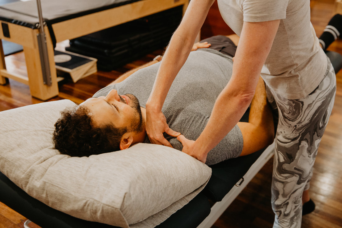
Physiotherapy can play a significant role in the management and treatment of shoulder bursitis. Manual therapy — hands-on techniques including massage, joint mobilisations and soft tissue manipulation — can help improve joint mobility, reduce muscle tension and enhance circulation in the affected area. Range of motion exercises maintain or improve shoulder joint mobility. Strengthening exercises targeting the muscles around the shoulder, including the rotator cuff muscles, improve strength and stability. Posture correction addresses how postural factors contribute to shoulder bursitis.
Rotator cuff strengthening — specifically targeting the external rotators (infraspinatus and teres minor) and subscapularis — restores the dynamic depression of the humeral head that is the primary mechanism protecting the subacromial space. Scapular stabiliser strengthening — lower trapezius, serratus anterior and middle trapezius — restores the scapular control that elevates the acromion during arm elevation, dynamically increasing the subacromial space. This combination is the most important and most evidence-based exercise intervention for subacromial bursitis.
Real time ultrasound assists in retraining rotator cuff activation where pain and guarding have disrupted normal neuromuscular patterns.
Dry needling of the periscapular muscles — upper trapezius, levator scapulae — addresses the secondary muscle tension that contributes to poor scapular control and elevated shoulder pain.
Thoracic spine mobilisation addresses the thoracic kyphosis component — improving thoracic extension directly improves scapular position and subacromial space. Postural and ergonomic education — workstation setup, sleeping position, activity modification — addresses the environmental contributors that will perpetuate bursitis if not modified alongside clinical treatment.
Clinical Pilates provides an excellent controlled environment for progressive rotator cuff and scapular strengthening. For patients whose shoulder bursitis developed in a workplace context, WorkCover funded physiotherapy is available.
For the post-surgical pathway following subacromial decompression, see our dedicated surgical rehabilitation page.
Our physiotherapists Bethany Kippen and Emma Cameron both have experience in shoulder conditions and are members of the Australian Physiotherapy Association.
To book or find out more, call us on 07 3706 3407 or book online below. We see patients from across Brisbane's southside including Tarragindi, Coorparoo, Holland Park, Greenslopes and Mt Gravatt.
Shoulder bursitis is inflammation of one of the bursae around the shoulder joint — most commonly the subacromial bursa, which sits between the rotator cuff tendons and the undersurface of the acromion (the bony roof of the shoulder). When this bursa becomes irritated or inflamed it fills with excess fluid, producing pain, swelling and restricted shoulder movement that is typically worst with overhead activity, reaching and lying on the affected side.
Shoulder bursitis is one of the most common causes of shoulder pain in adults — the subacromial space is a narrow anatomical corridor and any factor that reduces its dimensions or increases the load on the structures within it can produce bursal irritation. It is closely related to shoulder impingement syndrome and rotator cuff tendinopathy — in practice these conditions frequently coexist, and differentiating between them requires a careful clinical assessment rather than assuming the imaging diagnosis tells the whole story.
The subacromial space — why it matters
The subacromial space is the gap between the humeral head below and the acromion and coracoacromial ligament above, through which the supraspinatus tendon and subacromial bursa must pass during shoulder elevation. In a healthy shoulder, the rotator cuff muscles — particularly the supraspinatus — depress the humeral head during elevation, maintaining adequate subacromial space. When rotator cuff function is compromised from weakness, fatigue or inhibition, the humeral head rides higher in the socket, narrowing the subacromial space and increasing mechanical contact on the bursa and tendon.
This dynamic narrowing is the central mechanism of most subacromial bursitis presentations — it is a functional problem driven by rotator cuff and scapular muscle imbalance, not simply an anatomical problem with the shape of the acromion. This understanding is clinically important because it means physiotherapy addressing rotator cuff function and scapular control directly addresses the mechanism driving the bursitis — not just managing the symptoms.
What causes shoulder bursitis?
Several factors contribute to subacromial bursitis. Rotator cuff weakness or dysfunction — from overuse, injury or disuse — reduces the dynamic depression of the humeral head during shoulder movement. Poor scapular control — weak lower trapezius and serratus anterior — produces inadequate acromial elevation during shoulder elevation, reducing the subacromial space. Repetitive overhead activity in occupational or sporting contexts — painting, swimming, throwing, racquet sports — creates sustained mechanical loading on the bursa.
Postural factors — forward head posture, thoracic kyphosis, rounded shoulders — alter shoulder mechanics and reduce subacromial space.
Inflammatory conditions including rheumatoid arthritis and crystal arthropathy directly cause bursal inflammation. Acromioclavicular joint osteoarthritis produces inferior osteophytes that mechanically impinge on the subacromial contents. Direct trauma — a fall onto the outstretched hand or onto the point of the shoulder — can cause acute bursal haemorrhage and inflammation.
What are the symptoms?
Pain that worsens with overhead movements or reaching is the hallmark. A painful arc — where shoulder elevation is comfortable from 0 to 60 degrees, painful from 60 to 120 degrees, and comfortable again above 120 degrees — is the classic clinical finding of subacromial impingement and bursitis. Pain is typically located in the lateral deltoid region and anterior shoulder, and is often worse at night, particularly when lying on the affected side.
Stiffness and weakness with sustained overhead or reaching activities, and pain with reaching across the body — the impingement test — are consistent features. Referred pain into the lateral arm is common from both the inflamed bursa and the associated rotator cuff tendinopathy.
How is it diagnosed?
Diagnosis of shoulder bursitis typically involves a physical examination by a healthcare provider, along with a review of your medical history. Imaging tests like X-rays or ultrasound may be used to rule out other shoulder conditions.
Clinical assessment includes the Neer and Hawkins-Kennedy impingement tests, rotator cuff strength testing, scapular assessment, and the painful arc assessment. Ultrasound directly visualises bursal fluid and thickening, identifies rotator cuff tears, and can guide diagnostic or therapeutic corticosteroid injection. MRI provides the most comprehensive assessment of the rotator cuff, bursa and surrounding structures.
A diagnostic injection of local anaesthetic into the subacromial space — producing temporary relief of symptoms — confirms the subacromial origin of pain and assists in differentiating bursitis from glenohumeral joint or acromioclavicular joint sources.
How can physiotherapy help?
Physiotherapy is the primary treatment for subacromial bursitis and addresses both the acute inflammation and the underlying biomechanical contributors that produced and perpetuate it.
In the acute phase, activity modification — temporarily avoiding the overhead and reaching activities that most provoke the bursa — reduces the repetitive mechanical irritation while the bursa settles. Ice application and relative rest from provocative activities manage symptoms without the deconditioning that complete rest produces.
Physiotherapy can play a significant role in the management and treatment of shoulder bursitis. Manual therapy — hands-on techniques including massage, joint mobilisations and soft tissue manipulation — can help improve joint mobility, reduce muscle tension and enhance circulation in the affected area. Range of motion exercises maintain or improve shoulder joint mobility. Strengthening exercises targeting the muscles around the shoulder, including the rotator cuff muscles, improve strength and stability. Posture correction addresses how postural factors contribute to shoulder bursitis.
Rotator cuff strengthening — specifically targeting the external rotators (infraspinatus and teres minor) and subscapularis — restores the dynamic depression of the humeral head that is the primary mechanism protecting the subacromial space. Scapular stabiliser strengthening — lower trapezius, serratus anterior and middle trapezius — restores the scapular control that elevates the acromion during arm elevation, dynamically increasing the subacromial space. This combination is the most important and most evidence-based exercise intervention for subacromial bursitis.
Real time ultrasound assists in retraining rotator cuff activation where pain and guarding have disrupted normal neuromuscular patterns.
Dry needling of the periscapular muscles — upper trapezius, levator scapulae — addresses the secondary muscle tension that contributes to poor scapular control and elevated shoulder pain.
Thoracic spine mobilisation addresses the thoracic kyphosis component — improving thoracic extension directly improves scapular position and subacromial space. Postural and ergonomic education — workstation setup, sleeping position, activity modification — addresses the environmental contributors that will perpetuate bursitis if not modified alongside clinical treatment.
Clinical Pilates provides an excellent controlled environment for progressive rotator cuff and scapular strengthening. For patients whose shoulder bursitis developed in a workplace context, WorkCover funded physiotherapy is available.
For the post-surgical pathway following subacromial decompression, see our dedicated surgical rehabilitation page.
Our physiotherapists Bethany Kippen and Emma Cameron both have experience in shoulder conditions and are members of the Australian Physiotherapy Association.
To book or find out more, call us on 07 3706 3407 or book online below. We see patients from across Brisbane's southside including Tarragindi, Coorparoo, Holland Park, Greenslopes and Mt Gravatt.
Who to book in with:
Yulia Khasyanova
|
Emma Cameron
|
Bethany Kippen
|