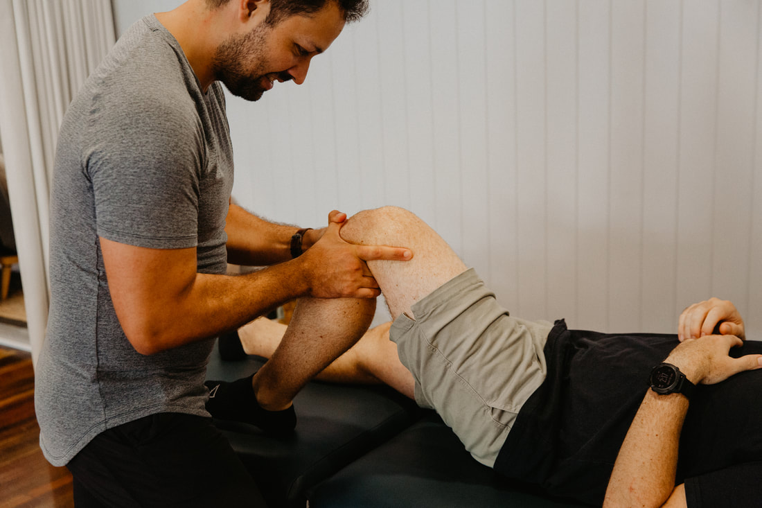
Knee dislocation physiotherapy Brisbane southside.
What is a Knee Dislocation?
A knee dislocation is a serious injury that occurs when the bones that form the knee joint are forced out of position. This can happen as a result of a traumatic injury, such as a car accident or a sports injury. Knee dislocations are rare but can be very damaging to the knee and surrounding tissues.
How is a Knee Dislocation Diagnosed?
A knee dislocation is usually diagnosed through a combination of physical examination, imaging tests such as X-rays or MRI, and evaluation of symptoms. The doctor will look for signs of swelling, bruising, and instability in the knee joint. They will also test the range of motion of the knee joint and look for any abnormal movements or pain.
How Can Physiotherapy Help After a Knee Dislocation?
Physiotherapy plays a crucial role in the rehabilitation of knee dislocations. The physiotherapist will work with the patient to develop a personalised treatment plan that includes exercises to help strengthen the muscles around the knee joint. They will also teach the patient how to safely perform activities of daily living, such as walking and standing, without putting too much strain on the injured knee.
In addition to exercises, the physiotherapist may also use other modalities such as ultrasound, heat or cold therapy, and electrical stimulation to help reduce pain and inflammation in the knee joint. They may also provide manual therapy, such as massage or joint mobilisation, to help improve mobility and flexibility in the knee joint.
How Can Clinical Pilates Help With Knee Dislocation Rehabilitation?
Clinical Pilates is a form of exercise that focuses on strengthening the core muscles of the body, including the muscles around the knee joint. Clinical Pilates can be an effective tool for knee dislocation rehabilitation as it helps to improve stability and balance in the knee joint, reducing the risk of further injury.
The physiotherapist will work with the patient to develop a Pilates program that is tailored to their specific needs and goals. The Pilates exercises will be designed to help improve core strength, balance, and flexibility, all of which are important for knee dislocation rehabilitation.
What Is the Prognosis for Knee Dislocation?
The prognosis for knee dislocation depends on the severity of the injury and the treatment that is received. In general, knee dislocations are considered to be serious injuries that require prompt medical attention and rehabilitation.
With appropriate treatment, most people with knee dislocations are able to regain function and return to their normal activities. However, it can take several months of rehabilitation and physiotherapy before the knee is fully healed and functional again.
Surgical Treatment for Knee Dislocations
When a knee dislocation is severe and has caused significant damage to the bones, ligaments, and other structures within the knee joint, surgical intervention may be required to restore proper alignment and function to the joint. The decision to undergo surgery will depend on a variety of factors, including the extent of the injury, the patient's overall health and age, and their goals for recovery.
The surgical approach will depend on the specific injuries sustained during the dislocation. Common surgical interventions include repairing or reconstructing torn ligaments, repositioning and stabilising the bones of the knee joint, or repairing any damaged soft tissues. The specific surgical approach will vary depending on the patient's individual needs and the extent of the damage.
After surgery, the patient will undergo a period of rehabilitation to help them regain strength, flexibility, and range of motion in the knee joint. The physiotherapist will work closely with the surgeon to develop a rehabilitation plan that is tailored to the patient's individual needs. This plan may include exercises to improve muscle strength and flexibility, manual therapy to improve joint mobility, and modalities such as heat or cold therapy to reduce pain and inflammation.
Long-Term Management of Knee Dislocations
Even after the knee has healed from a dislocation, there may be some long-term effects of the injury that can impact the patient's quality of life. Some patients may experience ongoing pain or stiffness in the knee joint, while others may be at increased risk for developing arthritis in the joint over time.
The physiotherapist can work with the patient to develop a long-term management plan that is designed to help them manage these symptoms and maintain their function and mobility. This may include ongoing exercises to help strengthen and stabilise the knee joint, as well as modalities such as heat or cold therapy to help manage pain and inflammation.
Additionally, the physiotherapist may work with the patient to develop strategies for preventing future injuries to the knee joint. This may involve education about proper body mechanics during physical activity, as well as strategies for managing pain or stiffness if it does occur.
In some cases, ongoing physiotherapy or other forms of treatment may be necessary to help the patient maintain their function and mobility over time. The physiotherapist can work with the patient to develop a long-term plan that is designed to meet their individual needs and goals for recovery.
Have you or a loved one recently suffered a knee dislocation? Our physiotherapists would love to help! Call our friendly clinic a call on 07 3706 3407, email [email protected] or make your initial appointment booking via the link below.
A knee dislocation is a serious injury that occurs when the bones that form the knee joint are forced out of position. This can happen as a result of a traumatic injury, such as a car accident or a sports injury. Knee dislocations are rare but can be very damaging to the knee and surrounding tissues.
How is a Knee Dislocation Diagnosed?
A knee dislocation is usually diagnosed through a combination of physical examination, imaging tests such as X-rays or MRI, and evaluation of symptoms. The doctor will look for signs of swelling, bruising, and instability in the knee joint. They will also test the range of motion of the knee joint and look for any abnormal movements or pain.
How Can Physiotherapy Help After a Knee Dislocation?
Physiotherapy plays a crucial role in the rehabilitation of knee dislocations. The physiotherapist will work with the patient to develop a personalised treatment plan that includes exercises to help strengthen the muscles around the knee joint. They will also teach the patient how to safely perform activities of daily living, such as walking and standing, without putting too much strain on the injured knee.
In addition to exercises, the physiotherapist may also use other modalities such as ultrasound, heat or cold therapy, and electrical stimulation to help reduce pain and inflammation in the knee joint. They may also provide manual therapy, such as massage or joint mobilisation, to help improve mobility and flexibility in the knee joint.
How Can Clinical Pilates Help With Knee Dislocation Rehabilitation?
Clinical Pilates is a form of exercise that focuses on strengthening the core muscles of the body, including the muscles around the knee joint. Clinical Pilates can be an effective tool for knee dislocation rehabilitation as it helps to improve stability and balance in the knee joint, reducing the risk of further injury.
The physiotherapist will work with the patient to develop a Pilates program that is tailored to their specific needs and goals. The Pilates exercises will be designed to help improve core strength, balance, and flexibility, all of which are important for knee dislocation rehabilitation.
What Is the Prognosis for Knee Dislocation?
The prognosis for knee dislocation depends on the severity of the injury and the treatment that is received. In general, knee dislocations are considered to be serious injuries that require prompt medical attention and rehabilitation.
With appropriate treatment, most people with knee dislocations are able to regain function and return to their normal activities. However, it can take several months of rehabilitation and physiotherapy before the knee is fully healed and functional again.
Surgical Treatment for Knee Dislocations
When a knee dislocation is severe and has caused significant damage to the bones, ligaments, and other structures within the knee joint, surgical intervention may be required to restore proper alignment and function to the joint. The decision to undergo surgery will depend on a variety of factors, including the extent of the injury, the patient's overall health and age, and their goals for recovery.
The surgical approach will depend on the specific injuries sustained during the dislocation. Common surgical interventions include repairing or reconstructing torn ligaments, repositioning and stabilising the bones of the knee joint, or repairing any damaged soft tissues. The specific surgical approach will vary depending on the patient's individual needs and the extent of the damage.
After surgery, the patient will undergo a period of rehabilitation to help them regain strength, flexibility, and range of motion in the knee joint. The physiotherapist will work closely with the surgeon to develop a rehabilitation plan that is tailored to the patient's individual needs. This plan may include exercises to improve muscle strength and flexibility, manual therapy to improve joint mobility, and modalities such as heat or cold therapy to reduce pain and inflammation.
Long-Term Management of Knee Dislocations
Even after the knee has healed from a dislocation, there may be some long-term effects of the injury that can impact the patient's quality of life. Some patients may experience ongoing pain or stiffness in the knee joint, while others may be at increased risk for developing arthritis in the joint over time.
The physiotherapist can work with the patient to develop a long-term management plan that is designed to help them manage these symptoms and maintain their function and mobility. This may include ongoing exercises to help strengthen and stabilise the knee joint, as well as modalities such as heat or cold therapy to help manage pain and inflammation.
Additionally, the physiotherapist may work with the patient to develop strategies for preventing future injuries to the knee joint. This may involve education about proper body mechanics during physical activity, as well as strategies for managing pain or stiffness if it does occur.
In some cases, ongoing physiotherapy or other forms of treatment may be necessary to help the patient maintain their function and mobility over time. The physiotherapist can work with the patient to develop a long-term plan that is designed to meet their individual needs and goals for recovery.
Have you or a loved one recently suffered a knee dislocation? Our physiotherapists would love to help! Call our friendly clinic a call on 07 3706 3407, email [email protected] or make your initial appointment booking via the link below.