Medial Collateral Ligament (MCL) Injuries physiotherapy Brisbane southside.
What is a Medial Collateral Ligament Injury?
A medial collateral ligament (MCL) injury is a most common type of knee injury, typically resulting from a direct blow to the outside of the knee on a planted foot. The MCL is a thick band of tissue that runs along the inner edge of the knee and helps to stabilize it. When the MCL is injured, it can cause pain, swelling, and instability in the knee joint.
What are the grades of a Medial Collateral Ligament Injury?
MCL injuries are often classified into three grades based on their severity:
What are the symptoms of a Medial Collateral Ligament Injury?
Common symptoms of an MCL injury may include:
How is a Medial Collateral Ligament Injury diagnosed?
A healthcare professional will typically diagnose an MCL injury through a physical examination, which may include tests like the valgus stress test to assess the stability of the knee. In some cases, imaging studies like an MRI may be used to confirm the diagnosis and assess the severity of the injury, rule in or out the associated medial meniscus tear.
What is the recovery time for a Medial Collateral Ligament Injury?
Recovery time varies depending on the severity of the injury and the chosen treatment. Mild MCL injuries may heal in a few weeks with conservative treatment, while severe injuries may take several months, especially if surgery is required.
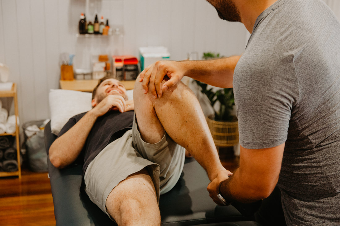
How can physiotherapy help a Medial Collateral Ligament Injury?
Physiotherapy plays a crucial role in the management and rehabilitation of MCL injury. Physiotherapy can help individuals with MCL injuries in various ways, depending on the severity of the injury and the specific needs of the patient. Grades I and II most often are treated non-operatively with physiotherapy being the main mode of rehabilitation. Here's how physiotherapy can assist in the recovery process:
If you or a loved one has questions about a Medial Collateral Ligament Injury and how our physiotherapists might be able to help please call us on 07 3706 3407 or email [email protected]. We would love to work with you!
A medial collateral ligament (MCL) injury is a most common type of knee injury, typically resulting from a direct blow to the outside of the knee on a planted foot. The MCL is a thick band of tissue that runs along the inner edge of the knee and helps to stabilize it. When the MCL is injured, it can cause pain, swelling, and instability in the knee joint.
What are the grades of a Medial Collateral Ligament Injury?
MCL injuries are often classified into three grades based on their severity:
- Grade I: A mild injury involving slight stretching of the ligament.
- Grade II: A moderate injury with partial tearing of the ligament.
- Grade III: A severe injury with a complete tear of the ligament.
What are the symptoms of a Medial Collateral Ligament Injury?
Common symptoms of an MCL injury may include:
- Pain and tenderness on the inner side of the knee.
- Swelling and bruising around the knee joint.
- Instability or a feeling that the knee is "giving way."
- Limited range of motion, especially the difficulty bending the knee
How is a Medial Collateral Ligament Injury diagnosed?
A healthcare professional will typically diagnose an MCL injury through a physical examination, which may include tests like the valgus stress test to assess the stability of the knee. In some cases, imaging studies like an MRI may be used to confirm the diagnosis and assess the severity of the injury, rule in or out the associated medial meniscus tear.
What is the recovery time for a Medial Collateral Ligament Injury?
Recovery time varies depending on the severity of the injury and the chosen treatment. Mild MCL injuries may heal in a few weeks with conservative treatment, while severe injuries may take several months, especially if surgery is required.
How can physiotherapy help a Medial Collateral Ligament Injury?
Physiotherapy plays a crucial role in the management and rehabilitation of MCL injury. Physiotherapy can help individuals with MCL injuries in various ways, depending on the severity of the injury and the specific needs of the patient. Grades I and II most often are treated non-operatively with physiotherapy being the main mode of rehabilitation. Here's how physiotherapy can assist in the recovery process:
- Pain Management: Physiotherapists can use various modalities, such as ice, heat, or electrical stimulation, to help manage pain and reduce swelling in the early stages of injury.
- Range of Motion: Physiotherapists will work with patients to maintain or regain a full range of motion in the injured knee joint. Gentle exercises and manual therapy techniques can be used to prevent stiffness.
- Strength Training: Strengthening the muscles around the knee joint is a critical aspect of MCL injury rehabilitation. Physiotherapists will design a tailored exercise program to strengthen the quadriceps, hamstrings, calf muscles, and hip muscles. Strong muscles can provide better support to the knee joint.
- Balance and Proprioception: Balance and proprioception exercises are essential to help patients regain stability and prevent future injuries. These exercises challenge the body's ability to maintain balance and spatial awareness, which is crucial for knee joint stability.
- Gait Training: Physiotherapists can assess and correct any abnormalities in your walking or running patterns caused by the MCL injury. They may recommend using assistive devices like crutches or braces during the early stages of recovery.
- Functional Training: As the healing progresses, physiotherapists will focus on functional activities that mimic everyday movements and sports-specific activities. This helps patients regain the ability to perform their usual activities safely and effectively.
- Patient Education: Physiotherapists educate patients on proper body mechanics and techniques to prevent future injuries. They may also provide guidance on home exercises and self-care strategies.
- Progress Monitoring: Throughout the rehabilitation process, physiotherapists monitor the patient's progress and adjust the treatment plan as needed. This ensures that the rehabilitation program is tailored to the individual's specific needs and recovery pace.
- Return to Activity: Once the MCL has healed sufficiently, physiotherapists can assist in gradually reintroducing patients to their regular activities or sports. They provide guidance on when it is safe to return to full activity and how to minimse the risk of re-injury.
- Support and Motivation: Physiotherapists provide emotional support and motivation throughout the rehabilitation process, helping patients stay committed to their recovery goals.
If you or a loved one has questions about a Medial Collateral Ligament Injury and how our physiotherapists might be able to help please call us on 07 3706 3407 or email [email protected]. We would love to work with you!