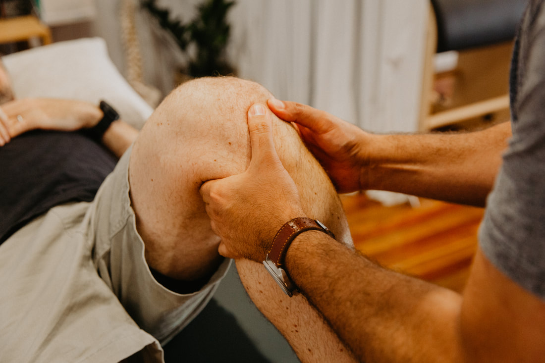
Knee Bursitis physiotherapy Brisbane southside.
What is a Knee Bursitis?
Bursitis of the knee is a condition that involves inflammation of the bursa sac located near the knee joint. Bursae are small, fluid-filled sacs that act as cushions between bones, tendons, muscles, and skin, reducing friction and allowing for smooth movement. When one or more of these bursae becomes inflamed, it can result in knee pain and discomfort.
What causes a Knee Bursitis?
The most common causes of knee bursitis include:
What are the symptoms of a Knee Bursitis?
Common symptoms of knee bursitis include:
What is the prevention for a Knee Bursitis?
To prevent knee bursitis, you can:
How can physiotherapy help with a Knee Bursitis?
Physiotherapy can play a crucial role in the management and rehabilitation of knee bursitis. A qualified physiotherapist can develop a personalized treatment plan to address the specific needs of the individual and help relieve pain, reduce inflammation, and improve knee function. Here's how physiotherapy can help with knee bursitis:
If you or a loved one has questions about Bursitis of the Knee and how our physiotherapists might be able to help please call us on 07 3706 3407 or email [email protected]. We would love to work with you!
Bursitis of the knee is a condition that involves inflammation of the bursa sac located near the knee joint. Bursae are small, fluid-filled sacs that act as cushions between bones, tendons, muscles, and skin, reducing friction and allowing for smooth movement. When one or more of these bursae becomes inflamed, it can result in knee pain and discomfort.
What causes a Knee Bursitis?
The most common causes of knee bursitis include:
- Repetitive Pressure: Frequent kneeling, as seen in occupations like carpet laying or gardening, can put excessive pressure on the prepatellar bursa, leading to inflammation.
- Direct Trauma: A blow or fall directly onto the kneecap can irritate and inflame the bursa.
- Infection: Although rare, bacterial infection of the bursa (septic bursitis) can occur, causing severe pain, swelling, and redness.
- Pes anserine bursitis, the inflammation of the bursa sac under the tendons on the inner side of knee is common in females with genu valgum, the increased knee-to-thigh angle, where additional pressure is exerted on the bursa due to anatomical angle of pelvis and thighs in females.
What are the symptoms of a Knee Bursitis?
Common symptoms of knee bursitis include:
- Pain and tenderness over the kneecap or the inner side of the knee
- Swelling and inflammation in the affected area
- Warmth and redness over the kneecap
- Difficulty kneeling or bending the knee.
- Limited range of motion
What is the prevention for a Knee Bursitis?
To prevent knee bursitis, you can:
- Use knee pads or cushions when kneeling for extended periods.
- Avoid repetitive kneeling or apply proper padding if necessary.
- Use proper kneeling techniques to reduce pressure on the knees.
- Maintain adequate muscle strength to allow good knee joint stability and articulation and reduce strain on the tendons and underlying bursa.
How can physiotherapy help with a Knee Bursitis?
Physiotherapy can play a crucial role in the management and rehabilitation of knee bursitis. A qualified physiotherapist can develop a personalized treatment plan to address the specific needs of the individual and help relieve pain, reduce inflammation, and improve knee function. Here's how physiotherapy can help with knee bursitis:
- Pain Management: Physiotherapists can use various techniques to alleviate pain, such as manual therapy, modalities like ice or heat, and therapeutic taping. They may also recommend over-the-counter or prescription pain medications if necessary.
- Reduce Inflammation: Physiotherapists can employ modalities like ultrasound or electrical stimulation to help reduce inflammation in the affected bursa.
- Range of Motion Exercises: Knee bursitis can lead to stiffness and limited range of motion. Physiotherapists can design exercises and stretches that target specific muscle groups to improve flexibility and restore normal range of motion in the knee joint.
- Strengthening Exercises: Weak muscles around the knee can contribute to bursitis. A physiotherapist will create a strengthening program to target the muscles that support the knee joint. Strengthening these muscles can help stabilize the knee and prevent future episodes of bursitis.
- Functional Rehabilitation: Physiotherapists work with patients to improve their ability to perform daily activities and functional tasks without pain. They may use functional exercises that mimic real-life movements to help patients regain their independence.
- Posture and Biomechanics: Poor posture and faulty biomechanics can contribute to knee bursitis. Physiotherapists can evaluate your posture and movement patterns and provide guidance on proper body mechanics to reduce stress on the knee joint.
- Manual Therapy: Hands-on techniques, such as joint mobilization and soft tissue massage, can be used by physiotherapists to reduce muscle tightness, improve circulation, and promote healing.
- Education: Physiotherapists educate patients about their condition, including its causes and contributing factors. They also provide guidance on self-management strategies, such as home exercises and ergonomic changes.
- Progressive Rehabilitation: As the patient's condition improves, physiotherapists adjust the treatment plan to gradually increase the intensity and complexity of exercises, promoting a safe return to full function.
- Prevention: Physiotherapists can educate patients on techniques and strategies to prevent future episodes of knee bursitis, such as proper warm-up and stretching routines, and advice on protective gear.
If you or a loved one has questions about Bursitis of the Knee and how our physiotherapists might be able to help please call us on 07 3706 3407 or email [email protected]. We would love to work with you!